Popular Press vs Scientific Journal Articles:
Insight Into the Color Blindness Gene and Gene Therapy
This web page was produced as an assignment for an undergraduate course at Davidson College.
Gene therapy for red-green color blindness in adult primates
In “Gene therapy for red-green colour blindness in adult primates,” Mancuso et al. attempt to restore trichromatic vision in squirrel monkeys missing the L opsin gene. The researchers did this by injecting a recombinant adeno-associated virus containing the L opsin gene into the photoreceptor layer of the monkey’s eyes. The researchers tested the success of the injections by training the monkeys to do a computer-based test called the Cambridge Color Test. They also tested to make sure the injected gene was producing its protein product by detecting GFP that was also injected into the eye using the same virus design. The only difference was that the GFP gene replaced the L opsin gene. The researchers then observed whether or not the monkeys had gained trichromatic vision by detecting spectral sensitivity using a wide-field color multifocal electroretinogram (mf-ERG) of their design (Mancuso et al. 2009).
Results: Here are the figures the researchers provided with and explanation of what was done.
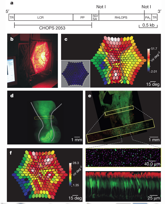
Figure 1: Part A of this figure shows the vector construct that was injected into the monkeys eyes. TR: terminal repeats, LCR: locus control region, PP: proximal promoter, SD/SA: splice donor/acceptor, RHLOPS: recombinant human L opsin cDNA, PA: polyadenylation signal. B,C, and F are mfERG readings for red light stimulus, 40 weeks after two injections of the two viruses (L opsin and GFP coding), and 70 weeks after three injections respectively. D shows a flourescence photo of the same area depicted in C. E was produced using confocal microscopy, green: GFP-expressing cells, magenta dots: cone locations, red: anti-M/L opsin antibody staining (Mancuso et al. 2009). Permission Pending*
Conclusions drawn from this figure: After injection of the viruses, it is clear from the mfERG readings that the monkey's eyes did produce middle to long wavelength absorbing cones (B, C, and E). Figure D shows the location of the subretinal injections and the gray lines depict the same area in figure C, showing that the largest amount of longer wavelength absorbing cones develop where the viruses were injected.
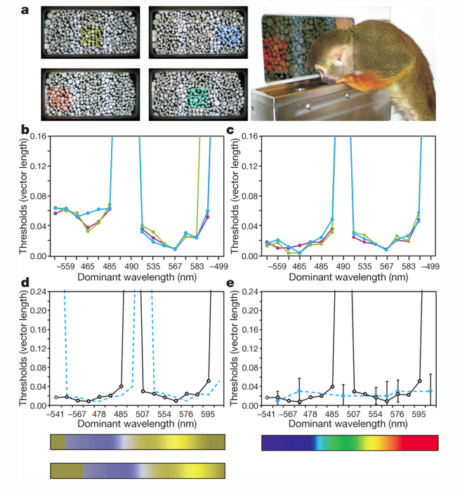
Figure 2: A is and example of the vision test the researchers administered to the monkeys. B shows results of the test for monkey 1 before gene therapy. C shows the results of the color vision test for monkey 2 before gene therapy. D shows the color spectrum of a dichromat. E shows a trichromat female control (dashed line) overlayed on D (Mancuso et al. 2009). Permission Pending*
Conclusions from Figure 2: Together, Figures2 B, C, and D show that the two monkeys are dichromat pre-treatment and the expected test outcomes if the monkeys were trichromat (E).
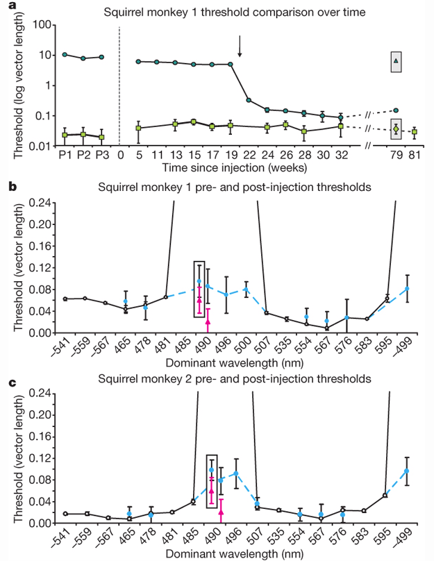
Figure 3: A shows the threshold for blue-green color confusion over time pre (p1-3) and post-treatment. B and C show a comparison between pre-treatment (solid)and post-treatment (blue dashed) thresholds for monkey 1 and 2 respectively. The pink triangles represent trichromatic female control thresholds (Mancuso et al. 2009). Permission Pending*
Conclusion from Figure 3: This figure shows that after twenty weeks the monkeys can differentiate between blue and green, indicating the adoption of trichromatic vision (A). The individual monkeys also exhibit trichromatic vector lengnth thresholds after gene therapy, once again indicating the gene therapy has worked (B and C).
Overall Conclusions and discussion in the paper
The researchers concluded from their data that, in the two monkeys they represented, gene therapy was sufficent to restore trichromatic vision to previously bichromatic individuals. They also talk about previous research claiming it is necessary that optical stimulation must be present durring development in order for the brain to detect it later in life. Their findings somwhat contradict this pervious assumption because monkeys that could not percieve the difference between blue and green light during development were able to percieve the difference after gene therapy. This leads the researchers to question the what is necessary to add a new color to one's vision (Mancuso et al. 2009).
Reference:
Mancuso, Katherine and Hauswirth, William W. and Li, Qiuhong and Connor, Thomas B. and Kuchenbecker, James A. and Mauck, Matthew C. and Neitz, Jay and Neitz, Maureen. 2009. Gene therapy for red–green colour blindness in adult primates. Nature. 461: 784-787
Full article can be found here:
http://www.nature.com/nature/journal/v461/n7265/full/nature08401.html
Genomics Page
Biology Home Page
Email Questions or Comments to Joholzwarth@davidson.edu
© Copyright 2011 Department of Biology, Davidson College, Davidson, NC 28035